
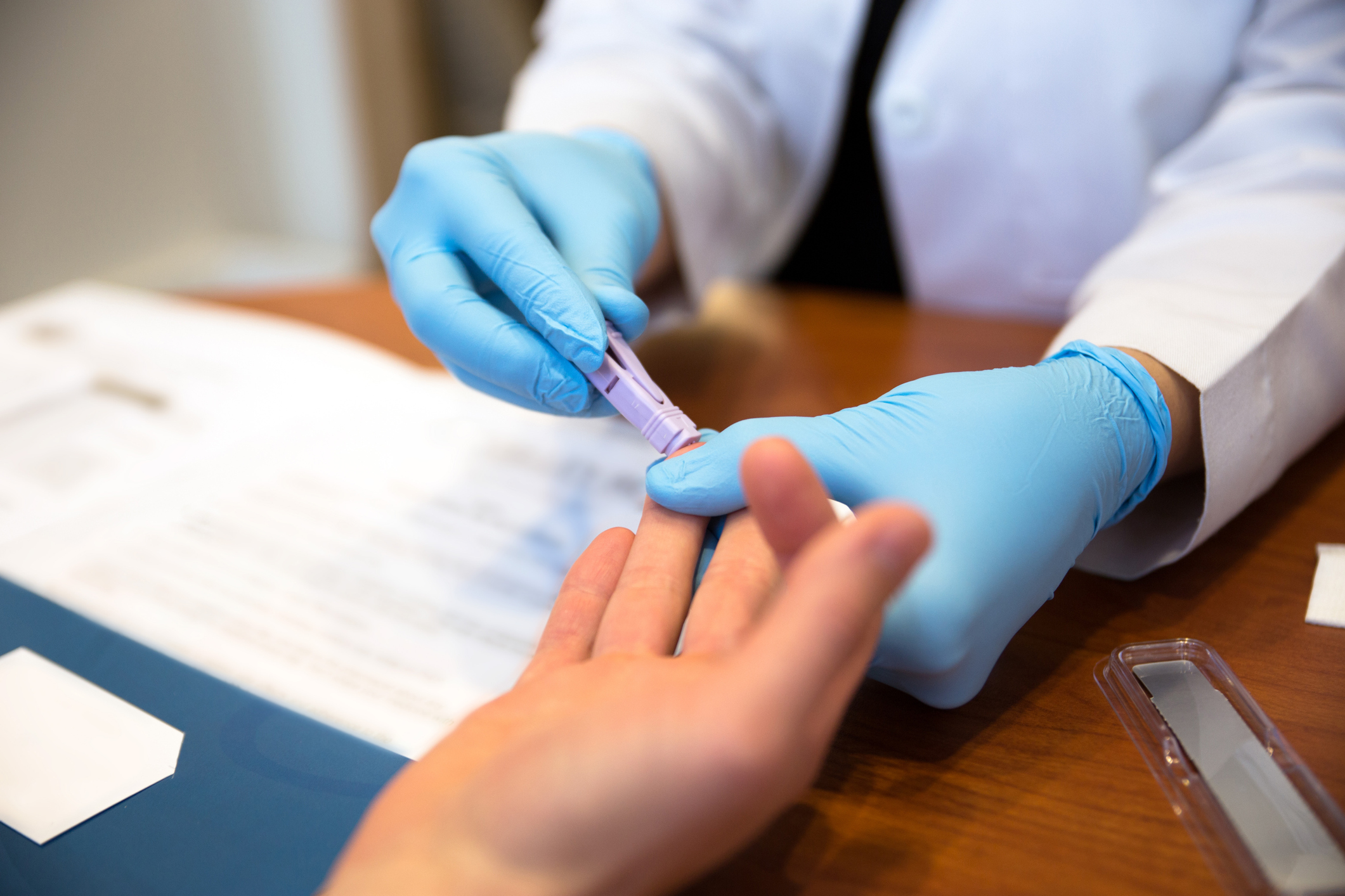
In vitro diagnostics (IVD) are diagnostic tests performed on biological samples (e.g. blood, tissue) obtained from the body to detect DNA/RNA, microorganisms, or biological biomolecules (e.g. protein) associated with diseases, infections or medical conditions. The IVD tests are generally non-invasive, used both in professional healthcare settings and at home as rapid kits and are useful for early detection of diseases, prevent the spread of diseases and improve patient care and management.

They are mainly used in point-of-care (POC) and laboratory settings. POC-based tests are very beneficial as it removes the need to send a sample to a laboratory for testing which reduces significantly the time between sample collection, especially useful in resource-poor settings where laboratories are located far away and there is a lack of good communication or transportation infrastructure. They include: blood glucose tests, COVID-19 tests, pregnancy tests and urinalysis test strips. Laboratory-based tests on the other hand require advanced equipment and expertise for complex medical conditions and include: blood grouping devices, human genetic tests, immunoassays and next-generation sequencing tests.
Prompt and effective therapeutic regimen is of vital importance in some public health infectious diseases such as Malaria with a rapid phase of general infection leading to death. Human malaria is caused by four types of Plasmodium parasites among which Plasmodium falciparum infection is the most fatal one. Upon infection, P. falciparum parasites go through two stages (pre-erythrocytic and intra-erythrocytic) of infecting Red Blood Cells (i-RBCs) in the human host which spread exponentially after only 48 hours and this necessitates the development of techniques for early diagnosis at low-rate infection of both early-stage (pre-erythrocytic) and late-stage (intra-erythrocytic) i-RBCs.
Various methods have been developed to separate i-RBCs from blood samples such as Giemsa-staining method, paper-based test kits, density gradient separation method, magnetic-activated cell separation (MACS). However, they are complex, well-trained personnel are required for reliable evaluation and have low accuracy at low infection rates.
Recently, a Magnetic IVD assay using magnetic separation to detect malaria has been developed. The method is effective at detecting the presence of the malaria parasite Plasmodium falciparum in patient serum, and it has the potential to be packed into a POC diagnostic tool. The technique does not require amplification and detection of the parasite via traditional culture methods or by PCR. It rapidly provides a diagnosis, and could potentially be developed into a compact package.
The technology depends on three basic technologies:
- Capture of Plasmodium falciparum DNA using sandwich hybridization
- Collection of the captured DNA using magnetic separation
- Detection of the captured DNA using ultra-bright nanorattles and Surface-enhanced Raman spectroscopy (SERS)
Capture of the malaria parasite DNA using sandwich hybridization
Sandwich hybridization is a single strand DNA or RNA capture technique whereby two oligonucleotide probes are synthesized, one complementary to the 5’ end and the other to the 3’ end. The probes can be attached to magnetic nanoparticles, fluorescent tags, polymer nanoparticles, or nanorattles to aid in the detection of the DNA or RNA of interest.
The technique discussed here uses magnetic nanoparticles to collect the captured DNA, and nanorattles to detect the DNA. One probe is conjugated to magnetic nanoparticles while the other probe is conjugated to nanorattles. If present, the DNA is hybridized between them, and links the magnetic particle and nanorattle together.
Collection of Plasmodium falciparum DNA using magnetic separation
After the sandwich hybridization incubation is complete, and the magnetic particle-DNA-nanorattle object is formed, there needs to be a way to collect it from serum and direct it to an area where it can be detected by SERS. This is accomplished by magnetic separation with the use of an external magnet. The magnetic nanoparticles are susceptible to the magnetic field, meaning that they become magnetized and move toward the magnet. The process is rapid, which is desirable in a POC diagnostic tool.
Detection of the captured DNA using ultra-bright nanorattles and SERS
Surface enhanced Raman spectroscopy is a non-fluorescent method of identifying molecules attached to metallic surfaces. Raman Scattering is a type of spectroscopy that measures information about the vibrational modes of molecules. A laser shines light onto the molecule, some light passes through, but some light is scattered. The Raman spectra are specific to each type of molecule, and when molecules are adsorbed to metallic surfaces the Raman scattering can be greatly enhanced. This technique has some advantages over fluorescence detection. These advantages include no bleaching, using a narrower light spectrum, and achieving a lower limit of detection. The nanorattle is a nanoparticle consisting of a gold core surrounded by SERS reporters contained by a silver shell coating. This ultra-bright nanorattle is very easy to detect and is therefore very useful in a diagnostic tool.
Since the Raman spectral lines for the ultrabritenanorattle are known it is easy to determine if the target is present in the sample or not. If the target DNA is not present then the magnetic particle-DNA-nanorattle sandwich is not made,and the nanorattles cannot be magnetically attracted to the measurement site. In that case no spectral lines are observed.
The system demonstrated a limit of detection of 3 picomolar, which means that it can detect Plasmodium falciparum DNA at concentrations as low as 10-10 M. The method has potential for development as a point of care in vitro diagnostic device for the detection of malaria.
The future of Malaria IVD assays
Given the above mentioned benefits of magnetic separation and the associating techniques, they are simple, sensitive, and suitable for automation, and look an attractive and promising procedure as portable platforms for POC molecular diagnostics which can highly and effectively increase the diagnosis of the Malaria at early phases and offer patients a much better therapeutic regimen.
Related news
- Detection of foodborne pathogens using magnetic separation
- Sandwich hybridization DNA/RNA capture using magnetic nanoparticles
- Using magnetic nanoparticles for molecular diagnosis of S. aureus