Our understanding of genetic material has substantially increased since Friederich Miescher first extracted DNA in 1869. He discovered that a material exists within cells that precipitates out of acidic solution and dissolves into alkaline solution. He called it nuclein because it seemed to be located within the nucleus. It took until 1953 for the structure of DNA to be elucidated. It was during this time that procedures to isolate DNA began to emerge. Later, during the 1960's and 70's scientists were furiously untangling the cellular environment, and the discovery of RNA with its various forms and functions further refined DNA purification procedures.
Free download: Magnetic DNA purification
The importance of DNA isolation ranges from research purposes to applications such as food development, identification, and more. DNA isolation allows clinicians to diagnose patients with genetic mutations, determining who is at high risk for cancer. DNA isolation in fetuses allows for determination of hereditary disorders, which allows for screening for certain mutations that can then be fixed with gene therapies. At pharmaceutical companies DNA isolation can be an initial step for developing targets for drugs. DNA isolation starts the process used for identifying paternity or family. DNA isolation can even be used to identify traits or characteristics that create more desirable fruit. DNA isolation allowed the Hawaiian pineapple industry single handedly to be saved from bacteria that nearly had the species go extinct. A DNA isolation that can be performed well is crucial to all these applications.
Early nucleic acid purification methods relied on density gradient separation. Later strategies such as guanidinium thiocyanate-phenol-chloroform extraction took advantage of the variable solubility of cellular components in organic or aqueous solvent. As our understanding of nucleic acid chemistry deepened, more researchers saw the benefit of isolating and studying genetic material, and new tools such as solid-phase centrifugation columns hit the market. These columns took advantage of selective binding of negatively charged nucleic acids mediated by salt concentration and solution pH. Recently, superparamagnetic nanoparticles have been introduced as solid-phase support systems for the purification of nucleic acids by magnetic separation.
There are three general steps to nucleic acid isolation
- Cell lysis
- Removal of contaminating proteins, nucleic acids, and salts and deactivating DNAases or RNAases.
- Recovery of DNA or RNA
Step 1: cell lysis
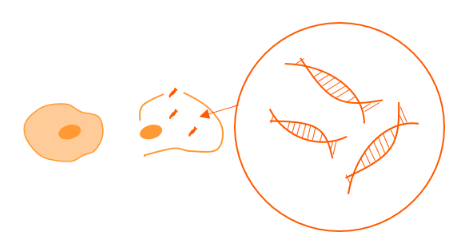
The first step in nucleic acid isolation is breaking apart the cell wall and/or membrane to release the genetic material. This is accomplished with the use of lysis buffer, rotor homogenizer, bead mill, freeze thaw cycles, or sonication. Lysis buffer contains a detergent to help break down cell membranes, and an enzyme such as protease K for digesting protein components. The homogenizer and bead mill provide rough mechanical shearing to disrupt the tissue and cells. Cell lysis produces a solution in which the cellular contents are no longer neatly compartmentalized. Therefore, DNAases and RNAases threaten to enzymatically destroy the genetic material. EDTA can be used to deactivate DNAases by chelating divalent ions that are necessary for enzymatic activity, and RNAases are permanently destroyed by beta-mercaptoethanol. Additionally, to decrease the chance of contamination with environmental RNAases or DNAases, it is important to use DEPC-treated water and buffers, to wear gloves, and to maintain a clean workspace.
Steps 2 and 3: Removal of contaminates and recovery of DNA and RNA
Guanidinium thiocyanate-phenol-chloroform extraction
This early purification technique takes advantage of the variable solubility of cellular components in organic or aqueous solvent, and sensitivity to salt concentration. The addition of phenol and chloroform to cell lysate causes the solution to fall out into a hydrophobic phase and a hydrophilic phase. Nucleic acids will remain in the hydrophilic phase while proteins will be in the hydrophobic phase. Guanidinium thiocyanate is a chaotropic agent that disrupts hydrogen-bonding. When guanidinium thiocyanate is used in a phenol-chloroform extraction it helps separate RNA and DNA into two different aqueous layers. After centrifugation the RNA will be dissolved in the top aqueous layer, with DNA below it, and the proteins will be in the organic hydrophobic layer at the bottom. Guanidinium thiocyanate also denatures proteins, including RNAases.
Ethanol precipitation and solid-phase column support systems
Nucleic acids can also be collected through ethanol precipitation methods. This technique requires solid phase support systems. The first support system developed for this purpose was a column. The column is static and the solution is poured through it. Under appropriate salt concentrations and pH the nucleic acids bind to the column while other contaminants flow through. This procedure relies on multiple washing and centrifugation steps to remove contaminants before finally eluting and capturing the DNA or RNA.
Magnetic nucleic acid purification
More recently, superparamagnetic particles and magnetic separation have been used to capture nucleic acids. This method enables free mobility for the particles within the solution, which improves nucleic acid adsorption and capture efficiency. The superparamagnetic nature of the particles allows them to be manipulated by an external magnet and retained in place while the contaminating proteins and salts are washed away. These systems can rely on salt concentration and ethanol precipitation or they can use more sophisticated chemistry for the reversible-binding of DNA or RNA in a specific or nonspecific manner.
Applications of Magnetic Nucleic Acid Purification
Much of nucleic acid isolation relies on the ability to distinguish genetic material from other genetic material. For example, cancerous cells exist alongside healthy cells in a human body, and distinguishing between the two is paramount to the diagnosis of cancer in an individual. DNA microarrays are a common application to distinguish between healthy and cancerous cells. Cells are grown, genetic material is isolated, and the material is then labeled with a fluorescent probe that binds to specific DNA sequences. Purification is highly important for this step, as the removal of salts and other chemicals used in isolation can interfere with fluorescent binding.
Related news